This 24-year-old reports for a knee MRI. She reports no injury, but her knee is misaligned and pain is worse with activity.
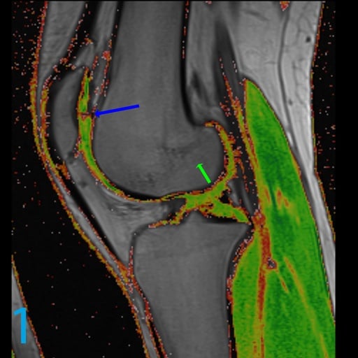
1. What do you call the type of image shown on image 1?
2. What parameter on MRI is varied to achieve such a map?
3. What is the name of the cardinal feature of cartilage organization that is appreciated on such a map?
4. What does the blue arrow indicate on image 1?
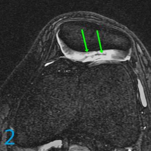
5. What adjectives might you use to describe the abnormality highlighted by the green arrows on image 2?
1. T2 cartilage map
2. The echo time or TE
3. Stratification. In other words, the deeper portions of cartilage are normally darker on water-weighted images and get lighter as one proceeds more superficially and this translates into a change in color from darker colors and hues to lighter colors and hues. This should remain organized throughout the young knee.
4. Alteration in glycosaminoglycans substance, arrangement, and altered proteogylcan milieu.
5. Intrasubstance delamination and intrasubstance separation of the middle layer of lateral patellar facet cartilage.
For more case review, visit MRI Online.
[gravityform id="4" title="true" description="true"]