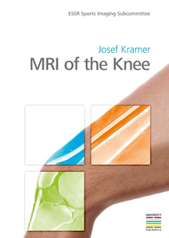
This 39-year-old man presents with a lump on his index finger and pain. He has a history of a pinching injury one year ago, so what would be your differential diagnosis of the mass indicated by the arrows? What would be your final diagnosis?
 Coronal T1
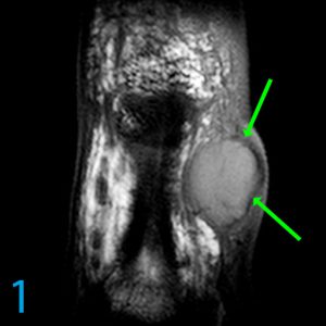
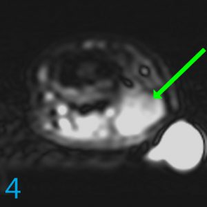
 PDW SPIR
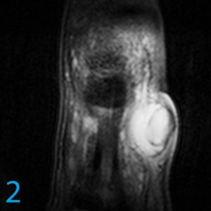
 Axial T1
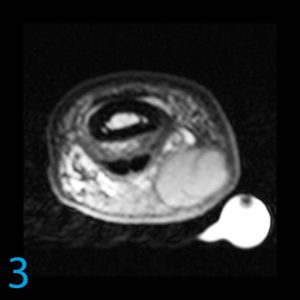
 Axial STIR
The following diagnoses should be considered in your differential diagnosis:
- Aneurysm or pseudoaneurysm: Although the signal on T1 is brighter than muscle, it is not the typical signal of blood, flow, clot, hemosiderin or thrombin. Pulsatile phase mismapping is not observed. Even though the location is reasonable for an aneurysm or pseudoaneurysm, the reasons mentioned make this diagnosis unlikely.
- Giant cell tumor of tendon sheath: This is one of the more common causes of a mass in the finger. However, giant cell tumor of tendon sheath has a very heterogeneous internal character "dusted" with hemosiderin and blood. These tend to be invasive and may even involve the bone even though they are benign. This lesion does not demonstrate those characteristics.
- Schwannoma: These are often cystic. However, they are not septated. Rather, they can exhibit small dotted foci of hypointensity as a manifestation of production of Antoni B cells within the schwannoma. As the digital nerve is in this proximity, the location is reasonable for a schwannoma. It would be unlikely, though, for a schwannoma to get this big before the patient came for evaluation. Schwannomas enhance, though no contrast was given in this case. Schwannomas of the finger are not typically associated with phakomatosis or NF2.
- Myxoid neoplasm or other tumor with myxoid character: Tough to exclude but singularly uncommon to rare in the finger.
- Epidermoid: Irregular, oval or round, intermediate T1, hyperintense T2, may be associated with trauma or penetrating trauma, internally septated. All of these findings are present in this case. Epidermoids demonstrate little to no enhancement, though no contrast was given.
This diagnosis in this case is a proven epidermoid. For more case review, check out MRI Online.
[gravityform id="4" title="true" description="true"]